
Immunohistochemistry
What we do
The Immunohistochemistry Platform is a biomedical research support platform. The main function is to offer biological sample processing services, both of human origin and animal models for analysis using histological techniques, from sample processing, sectioning and staining to digitization of preparations, histological analysis and results delivery. The platform offers the possibility of setting up new histological methods and stains, according to the demand and needs of our users, both internal and external. In addition, users receive training so they can use the platform's equipment in self-service mode.
Who We Are
Contact mail: immunohistoquimica@santpau.cat

Equipment
Immunohistochemistry Laboratory new IR-HSCSP building:
- ODYSSEY M. Image detection by chemiluminescence and near-infrared fluorescence. Membranes, such as Western Blot, Southern Blot and Northern Blot. Quantitative immunofluorescence. Cells in monolayer or suspension. Histological sections. Protease activity assays. Molecular imaging of mice. With Empiria Studio analysis software.

- SAKURA TISSUE TEK VIP 5 JR tissue processor

- KOS microwave tissue processor

- SAKURA TISSUE TEK TEC5 tissue embedding center with LEICA ARCADIA C Cold Plate

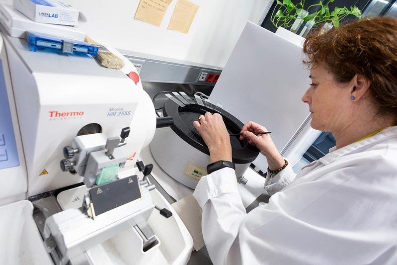
- MICROM HM355S rotary microtome with SELECTA TERMOFIN flotation bath and TERMARKS B8000 oven

- LEICA CM1950 cryostat.

- AGILENT Autostainer AS48 automatic immunostainer.

- SAKURA TISSUE TEK PRISMA automatic stainer (Sakura).

- GALILEO CK3500 semi-automatic tissue array.

- NUAIRE NU-S813-400E fume extraction hood

- Panoramic Scan II digital pathology scanner.

Immunohistochemistry Laboratory Pavilion 11
- LEICA EG1150H tissue embedding center: with operator and self-service

- LEICA EG1150C cold plate: with operator and self-service
- LEICA JUNG RM2055 microtome: with operator and self-service

- LEICA HI1210 flotation bath: with operator and self-service
- MEMMERT BE 300 oven

- LEICA JUNG CM3050S cryostat: with operator and self-service

- LEICA VT 1000S vibratome: with operator

- LABORSYSTEM fume extraction cabinet
- Panoramic MIDI BF digital pathology scanner

- Workstation for qualitative and quantitative histological analysis
Services
The Immunohistochemistry Platform offers the following services:
- Membrane detection, such as Western Blot, Southern Blot and Northern Blot. Quantitative immunofluorescence. Histological sections.
- Tissue decalcification.
- Tissue processing for paraffin and O.C.T. embedding.
- Paraffin and O.C.T. block sectioning.
- Fresh tissue sectioning.
- Antigen retrieval.
- Automatic, simple and double immunostaining.
- Stains: hematoxylin-eosin, giemsa, oil red, trichrome, PAS, Wright stain, alcian blue, safranin, sirius red, etc.
- Tissue microarray (TMA) preparation.
- Fluorescent in situ hybridization (FISH) on tissue.
- Digitization of histological preparations, under bright field, fluorescence and FISH conditions.
- Primary antibody optimization for conventional immunohistochemical staining and fluorescence.
- Histopathological and morphometric analysis of sections stained with routine histopathological and histochemical methods.
- Advice for research projects, theses and scientific papers.
The Platform offers the possibility of setting up new histological methods and stains, according to the demand and needs of our users, both internal and external.
Training
Some of the equipment used to carry out these techniques is accessible in self-service format for research staff after completing specific training.
The platform also provides other training courses or seminars on specific techniques or technologies related to genomics.
Quality
The Immunohistochemistry Platform has ISO 9001:2015 certification, which guarantees the quality of all the services it offers.
Rates
Check our rates.