
IR Sant Pau News
Stay up to date with the latest news from the RI
.png)
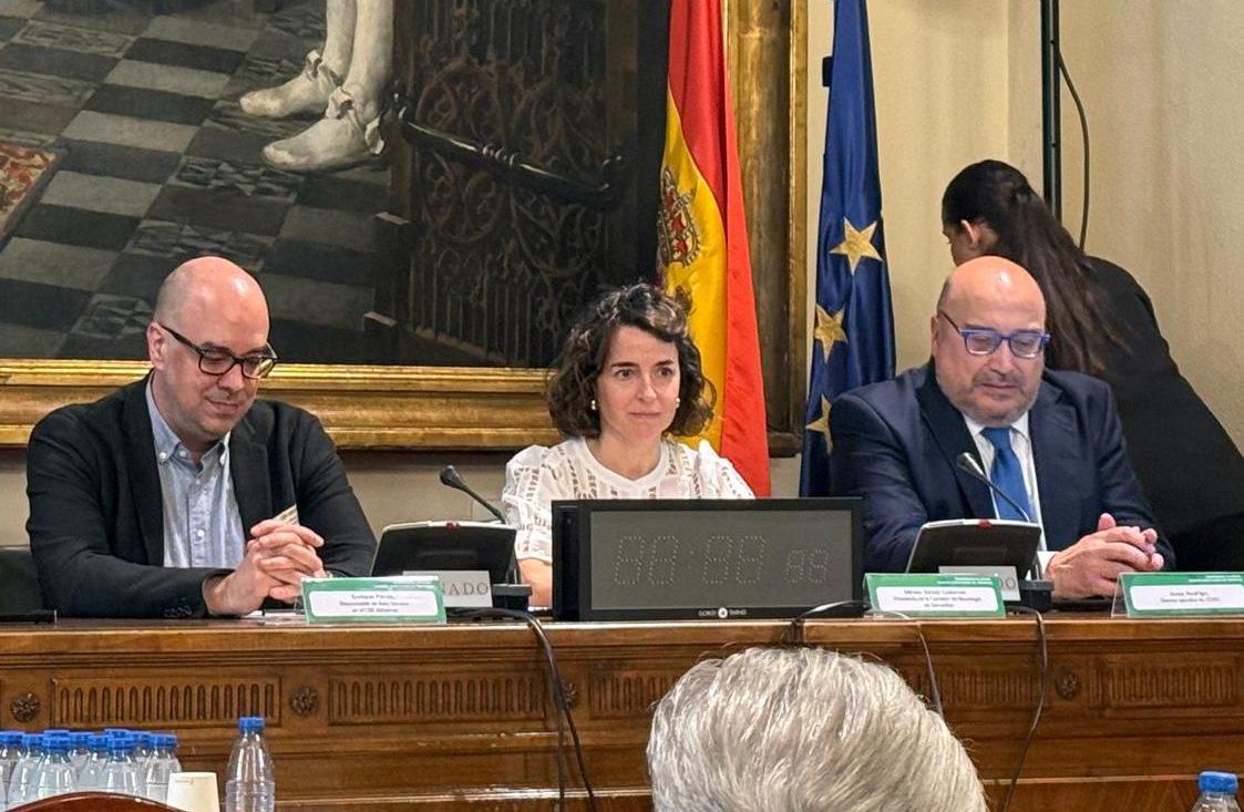
22/07/2026
Planetary Health Enters the International Clinical Guideline Development System for the First Time
.jpg)
.jpg)
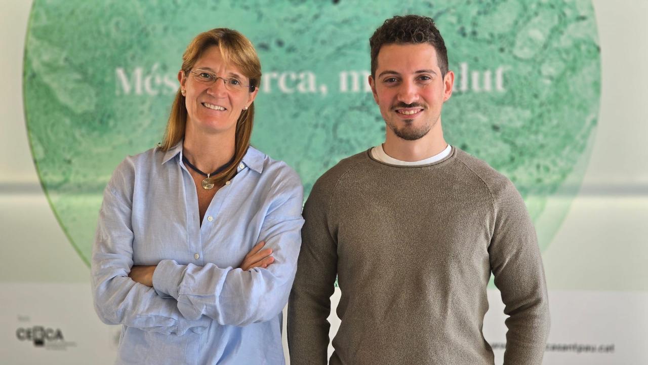
21/07/2026
Direct Study of Heart Tissue Opens New Avenues for Understanding Dilated Cardiomyopathy
.jpg)
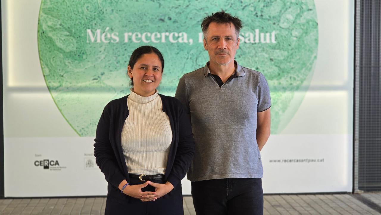
20/07/2026
Nanoligent, an IR Sant Pau Spin-Off, Recognized as a Catalonia Exponential Leader 2026
.jpg)
.jpg)
.jpeg)
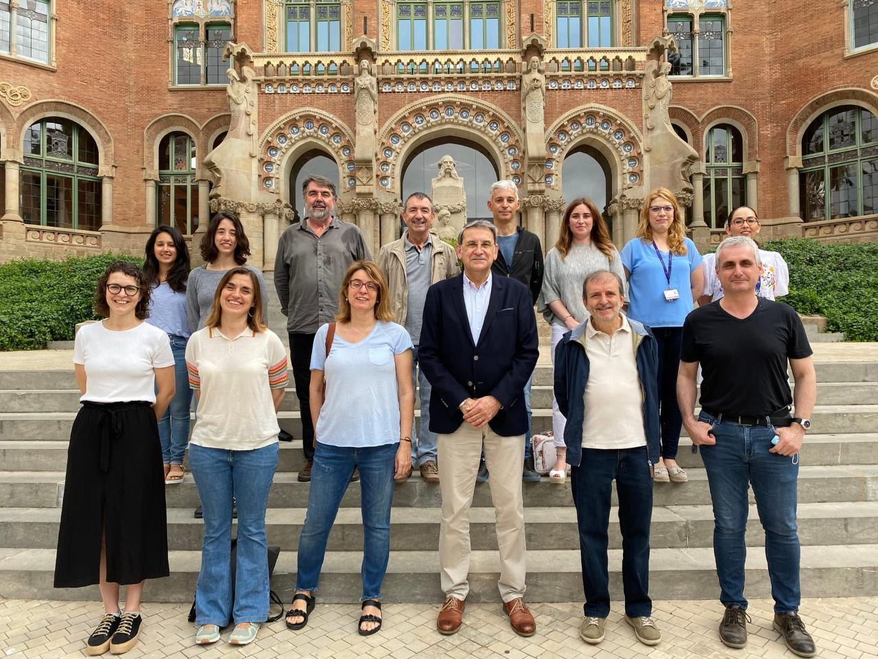
15/07/2026
IR Sant Pau Researchers Coordinate an International Special Issue on Lipid Metabolism, Cancer, and Cardiovascular Disease
.jpeg)
13/07/2026
Abatacept Reduces the Risk of Progression to Rheumatoid Arthritis in People With Palindromic Rheumatism
.jpg)
09/07/2026
A Fundación Mutua Madrileña Grant Will Support a Project on Pediatric Neuromuscular Diseases
.jpg)
07/07/2026
Researchers Develop an Artificial Intelligence–Based Tool Capable of Identifying Biological Profiles Associated With Thrombosis Risk
.jpg)
.jpg)
01/07/2026
Alzheimer’s Disease Biomarkers Can Predict Cognitive Decline in People Over 80 as Well
.jpg)
.jpg)
30/06/2026
Researchers Discover How Acute Myeloid Leukemia Invades the Lungs and Which Pathways Could Halt Its Infiltration
.jpeg)
29/06/2026
Familial Hypercholesterolemia Brings Together More Than 50 People at Sant Pau to Address Advances in Cardiovascular Prevention
25/06/2026
A Study Identifies Blood Biomarkers That Could Measure Response to Psychotherapy in Patients With Depression

18/06/2026
A SemFYC-Awarded Study Demonstrates the Value of Communication in Reducing Unnecessary Antibiotic Use in Children
17/06/2026
Researchers Discover a New Therapeutic Target to Prevent Thrombi With a Lower Bleeding Risk

10/06/2026
L’administració intravenosa d’atorvastatina durant l’infart redueix el dany miocàrdic en comparació amb l’administració d’una dosi de càrrega oral prèvia a l’infart
03/06/2026
Better Clinical Practice Guidelines Thanks to a New Method for Evaluating Multiple Treatments
Contact